
- Home
- About Us
- News
-
Research
- State Key Laboratory of High Performance Ceramics
- State Key Laboratory of Functional Crystals and Devices
- The Frontier Department
- Structural Ceramics and Composite Materials
- Information Functional Materials and Devices
- Inorganic Coating Materials
- Artificial Crystal
- Transparent Ceramics
- Energy Materials
- Biomaterials and Tissue Engineering
- Ancient Ceramics and Industrial Ceramics
- Pilot Test for New Materials
- People
- International Cooperation
- Education
- Journals
- Links
Scientists Realize the Osteochondral Regeneration via 3D Printed Bioceramic Scaffolds with Micro/Nanosize structured Surface
Osteochondral defect is a common disease in the clinic. Owning to the different physiological structures and functions of cartilage and subchondral bone, the regeneration of these two tissues remains a great challenge. In the previous studies, Prof. Chengtie Wu and Prof. Jiang Chang from Shanghai Institute of Ceramics, Chinese Academy of Sciences proposed the idea of using inorganic active ions to stimulate the regeneration of osteochondral defects. A series of 3D-printed bioceramic scaffolds with varied ionic components (Li, Mn, Sr, Si ions, etc.) were developed, and these scaffolds had been proved to have the capability to regenerate osteochondral defects effectively.
Recently, Prof. Wu and Prof. Chang put forward an innovative idea by harnessing the surface micro-nanostructure of 3D printed bioceramic scaffolds to regenerate the cartilage and subchondral bone, and important progress has been achieved.
Model bioactive biomaterials, bredigite (BRT) scaffolds, with controlled surface micro/nanostructure were successfully fabricated by combining 3D printing with a hydrothermal process. During the hydrothermal process, the complex surface of 3D porous scaffolds was uniformly contacted with the reaction solution; thus, the micro/nanostructured surface was homogeneously generated in the scaffolds. Furthermore, by using a hydrothermal method, micro/nanometer structures with highly controlled size and well-organized order could be achieved. Additionally, the growth of micro/nanostructured surface could reinforce the scaffolds by healing the microcracks. The in vitro studies indicated that micro/nanostructured surface significantly improved the adsorption of fibronectin (FN) protein, and promoted the adhesion, proliferation and differentiation of chondrocytes. Moreover, the activation of integrins α5β1 and αvβ1 in chondrocytes by micro/nanometer structure was, for the first time, observed. The mechanism of the micro/nanostructured surface in scaffolds promoting the differentiation of chondrocytes and regeneration of cartilage was elucidated. The micro/nanostructured surface first recruited FN protein from the surrounding environment, and then the adsorbed FN protein bound to integrin α and β subunits on cytomembrane and promoted clustering of integrins. Subsequently, the activated integrins facilitated F-actin remodeling and elevated the expression of cartilage specific genes (SOX9, Aggrecan, COL II, N-cadh), as well as maintained cell phenotype. Interestingly, the micro/nanostructured surface not only stimulated the differentiation of chondrocytes, but also significantly promoted the proliferation and differentiation of rBMSCs. It was found that micro/nanostructured surface in scaffolds significantly enhanced the early cell adhesion, and the cells displayed well-ordered F-actin. The expression of α5β1 and RhoA was significantly elevated, followed by stimulation of osteogenic differentiation markers (COL I, RUNX2, BMP2, OPN, and ALP). The results suggested that integrin α5β1 and RhoA coordinately regulated the fate of rBMSCs by mediating F-actin assembly, and further promoted osteogenic differentiation of rBMSCs. The in vivo studies revealed that the micro/nanostructured surface could significantly improve the reconstruction of both subchondral bone and cartilage. The results indicated that 3D printed scaffolds with micro/nanostructured surface can physiologically satisfy the demands for simultaneous reconstruction of both cartilage and subchondral bone, representing a smart strategy for the treatment of osteochondral defects. The paper has been published in Advanced Functional Materials (2018, 1806068, DOI: 10.1002/adfm.201806068.)
The related studies were supported by the National Key Research and Development Program of China, the National Natural Science Foundation of China and Key Research Program of Frontier Sciences CAS.

Micro/nano-structured surface in BRT scaffolds were successfully prepared via a hydrothermal process. In vitro, micro/nano-structured surface significantly promoted differentiation of chondrocytes via activating integrin α5β1 and integrin αvβ1, and stimulated osteogenic differentiation of rBMSCs through the synergetic effects of integrin α5β1 and RhoA. Furthermore, micro/nano-structured surface possessed dual physiological functions for the regeneration of both cartilage and subchondral bone.
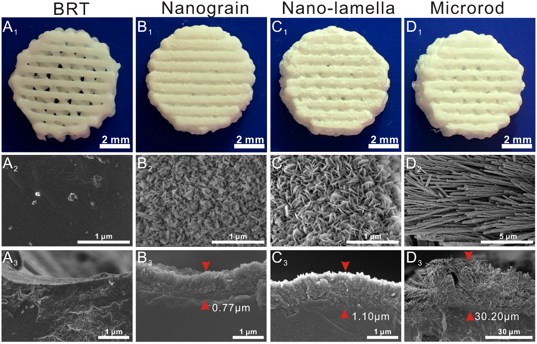
Morphology of BRT scaffolds before and after covering with micro/nanostructured surface. Photographs of A1) pure BRT scaffold, B1) nanograin, C1) nanolamella, and D1) microrod. SEM images of the surface of A2) pure BRT scaffold, B2) nanograin, C2) nanolamella, and D2) microrod. SEM images of cross section of A3) pure BRT scaffold, B3) nanograin, C3) nanolamella, and D3) microrod. The micro/nanostructured surface was uniformly generated in 3D scaffolds, and the thicknesses of nanograin, nanolamella, and microrod surfaces were ~0.77, ~1.10, and ~30.20 μm, respectively.
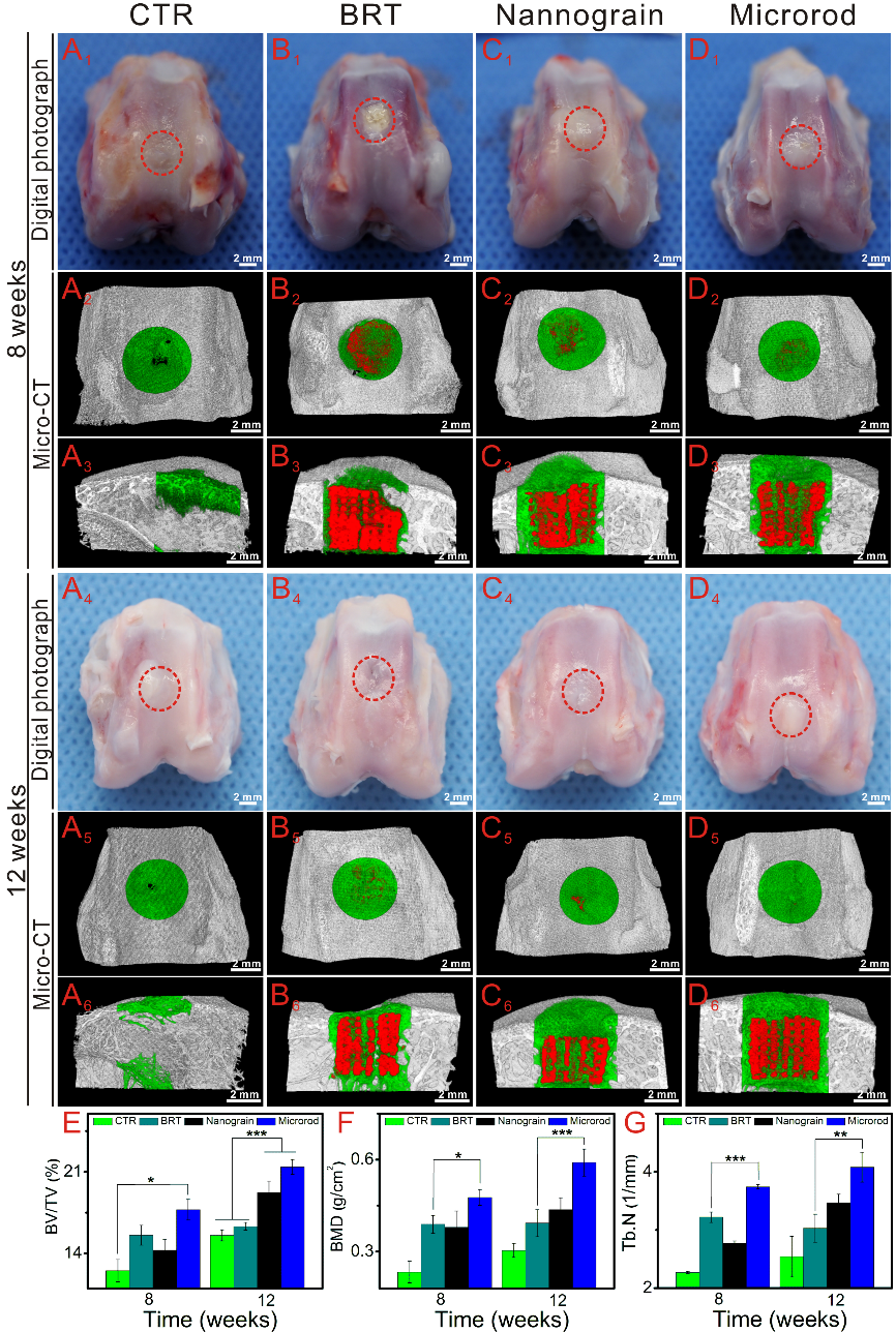
Photographs and micro-CT analysis of the defects at weeks 8 and 12 after surgery. Digital photographs of the defects at 8 and 12 weeks postsurgery: A1, A4) blank control without scaffolds, B1, B4) pure BRT scaffolds, C1, C4) nanograin, and D1, D4) microrod. A2–D2, A5–D5) The transverse view of micro-CT images at 8 and 12 weeks, respectively. A3–D3, A6–D6) The sagittal view of micro-CT images at 8 and 12 weeks, respectively. E) BV/TV, F) BMD, and G) Tb.N. In micro-CT images, red, green, and gray-white colors stand for the scaffold, new bone, and native bone, respectively. The
results of micro-CT analysis revealed that the new bone in the nanograin and microrod groups was obviously enhanced as compared to the CTR and BRT groups at both 8 and 12 weeks. Furthermore, the sagittal view of micro-CT images showed that the defects in the nanograin and microrod groups were filled with new bone, while in the CTR group new bone was only found in the margins of defects and large vacant space still existed in the middle of the defects. The bone assessment indicated that the microrod group had a greater level of bone formation and regeneration as compared to the CTR and BRT groups. Repeat number: n = 6 (*P < 0.05, **P < 0.01, ***P < 0.001).
Contact
Wu Chengtie
Shanghai Institute of Ceramics
E-mail: chengtiewu@mail.sic.ac.cn
Reference
Micro/Nanometer‐Structured Scaffolds for Regeneration of Both Cartilage and Subchondral Bone
https://onlinelibrary.wiley.com/doi/10.1002/adfm.201806068
