Strontium Ions Protect the Hearts Against Myocardial Ischemia/Reperfusion Injury
Myocardial infarction (MI) remains the major cause of morbidity and mortality worldwide. Timely restoration of blood supply following myocardial infarction is critical to save infarcted myocardium, while the reperfusion would cause additional damage.
The research group at Shanghai Institute of Ceramics of the Chinese Academy of Sciences tried to explore and develop optimal approaches with therapeutic and practical characters for the treatment of MI, especially, the simple and effective approaches aimed at protecting the heart against myocardial ischemia/reperfusion (I/R) injury.
Sr is an essential trace element in human body and Sr ions containing biomaterials are traditionally considered as a product for bone repair. They found out that Sr ions can promote the infarct heart recover from I/R injury. The result was published in Science Advances.
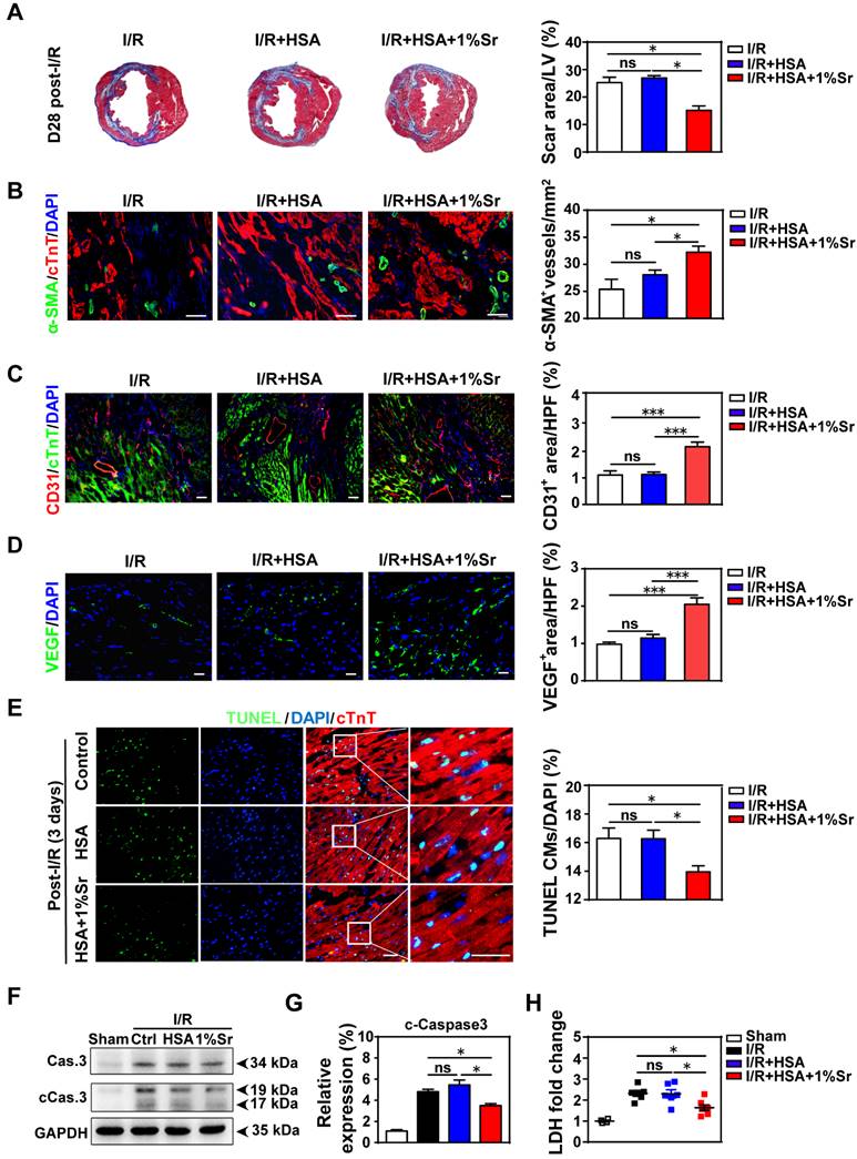
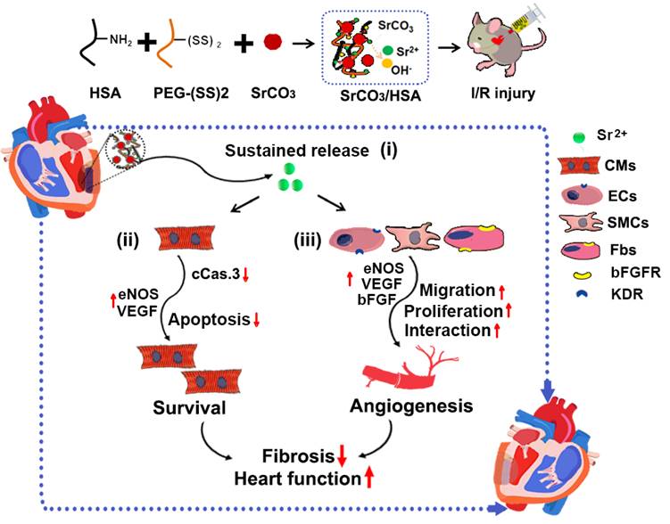
They reported that (i) Sr ions are indeed able to stimulate cellular activity of cardiomyocytes and blood vessel-forming cells; (ii) I/R-induced functional worsening and scar formation are significantly attenuated by the treatment with the Sr ions-containing composite hydrogels at the beginning of reperfusion; and (iii) these beneficial effects are accompanied with the reduced cardiomyocyte apoptosis and increased angiogenesis in the infarcted hearts.
The research demonstrates for the first time that Sr ions are able to protect cardiomyocytes and promote blood vessels activation. On this basis, the Sr ions activity for promoting cardiac repair of infarcted hearts is further confirmed by injection of designed SrCO3/HSA composite hydrogels in a murine I/R mode. Furthermore, in vitro cell experiments verify the beneficial effect of Sr ions on cardiomyocyte survival and vascular cells proliferation, migration and main angiogenesis related genes expression. Finally, reciprocal action between various cardiac cells, which related to blood vessel formation and maturation, are proved to be activated after cell co-culture and their cell-cell interaction can be promoted after Sr ions treatment through intercellular paracrine action.
All these findings suggest that Sr ions have the potential to be a therapeutic element for impaired cardiac tissues when locally applied in the infarcted myocardium, which may provide a new therapeutic strategy, partly with superior practicability due to the ions low-cost, stability, biosecurity and diversity, for the treatment of ischemic heart disease.
The research was supported by the National Key R&D Program of China (2016YFC1100201; 2017YFA0103700; 2016YFC1301204), the Strategic Priority Research Program of the CAS (XDA16010203; XDA16010201), the National Natural Science Foundation of China (32000945; 51902335; 81520108004, 81470422) and Shanghai Municipal Natural Science Foundation (19ZR1465200).
Reference:https://advances.sciencemag.org/content/7/3/eabe0726

SrCO3/HSA hydrogels inhibit myocardial fibrosis, increase angiogenesis and reduce cardiomyocyte apoptosis. (A) Representative cross-sectional images and quantitative data of scar area in LV on 5-μm slices stained with Masson’s trichrome at day 28 post-I/R; n = 5 hearts for each group. (B-D) Images and quantification of α-SMA+ blood vessels (B), CD31+ cells (C), and VEGF (D) in the border zone of infarcted hearts 28 days post-I/R. Scale bar = 50 μm, n = 4 hearts for each group. (E) Representative and quantification of staining for TUNEL+ cardiomyocytes in the border zone of infarcted hearts at day 3 post-I/R. Scale bar = 50 μm; n = 5-6 hearts for each group. (F-G) Representative and averaged western blot analysis for cleaved caspase-3 (c-Caspase3) and GAPDH in left ventricular heart tissues at day 3 post-I/R; n = 4 hearts each. (H) The concentrations of cardiac injury markers-LDH of the mice serum at day 3 post-I/R; n = 4-6 hearts each. Ctrl: I/R control, HSA: I/R + HSA, 1%Sr: I/R + HSA + 1%Sr, Cas3: Caspase3, cCas3: cleaved-Caspase3. All values were expressed as mean ± SEM; One-way ANOVA was used for statistical analyses. *P < 0.05, ***P < 0.001; ns: no statistical significance.

The schematic diagram of the SrCO3/HSA hydrogels in myocardium repair post-I/R. Various underlying mechanisms contributed to myocardial repair for SrCO3/HSA hydrogels post-I/R: (i) SrCO3/HSA hydrogels sustain the release of Sr ions in the infarct heat; (ii) Sr ions inhibit the apoptosis of CMs through reducing the active of caspase-3; (iii) Sr ions promote angiogenesis via increasing proliferation, strengthening paracrine capacity and stimulating the interaction of cardiac cells. Ultimately, alleviative fibrosis and elevated cardiac function are achieved after the treatment of SrCO3/HSA hydrogels. cCas3: cleaved-Caspase3; CMs: cardiomyocytes; ECs: endothelium cells; SMCs: smooth muscle cells; Fbs: fibroblasts.
Contact:
Prof. CHANG Jiang
Shanghai Institute of Ceramics
Email: jchang@mail.sic.ac.cn


