A progress in fabricating three-dimensional nanofibrous tubes with controllable micropatterns and macroscopic structures achieved by SIC-CAS
Electrospun ultrafine fibers with extremely long length and highly specific surface area have been found extensive applications in many biomedical and industrial fields. For example, electrospun fibrous tubes have shown a great potential in vascular, neural, and tendinous tissue engineering. Generally, rotating devices could be used to collect 3D fibrous tubes. Nevertheless, there still remains some disadvantages of this collecting method. For example, it is difficult to control the arrangements of fibers and the architectures of electrospun tubes. Meanwhile, failures in fabrication of tiny tubes (less than 0.3 mm), tubes with one closed end, and tubes with multiple interconnected tubular structures may also limit the applications of fibrous tubes. There exists considerable difficulties in fabricating fibrous tubes with controllable micropatterns and macroscopic 3D tubular structures synchronously, and the complex tissue structure and the fiber orientation still cannot be mimicked adequately.
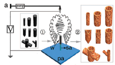
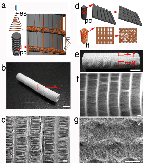
To overcome various limitations of the current preparation methods, Prof.Chang’s group of Shanghai Institute of Ceramics, Chinese Academy of Sciences has designed a unique static collecting method, which successfully fabricated nanofibrous tubes with different microscopic architectures and macroscopic 3D tubular structures. The novel method using novel 3D collecting templates is based on manipulation of electric field and electric forces. Micro and macro single tubes with multiple micropatterns, multiple interconnected tubes and many tubes with same or different sizes, shapes, structures and patterns can be prepared synchronously by using this unique technique. Parameters that could influence the order degree of patterned architectures have also been investigated. It is expected that electrospun tubes with controllable patterned architectures and 3D configurations may be attractive in many biomedical and industrial applications such as tissue engineering and filtration applications.
The related research work has recently been published in Nano Letters, 8(10), 3283-3287, 2008 (IF = 9.627), a leading journal in the field of nanoscale research, and a patent has been filed. This article was also selected as the Latest research highlights by Nature China. This work was financially supported by the National Basic Science Research Program of China (973 Program) (Grant 2005CB522704) and the Natural Science Foundation of China (Grant 30730034).
Related Links:
http://pubs.acs.org/cgi-bin/abstract.cgi/nalefd/2008/8/i10/abs/nl801667s.html
http://www.nature.com/nchina/2008/081008/full/nchina.2008.238.html


